Breast diagnostics
For mammograms (breast diagnostics), three different imaging techniques are available. Age, medical history and the issue involved are indicators for examination using one or more methods.
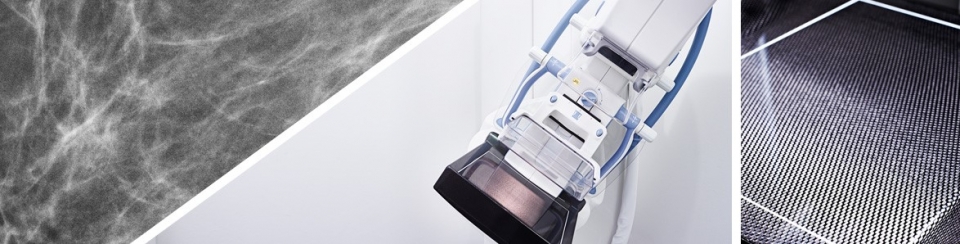
Digital mammography screening
Digital mammography screening is the main examination method used for breast diagnostics. It is used both for the early detection of breast cancer and the assessment of clinical symptoms.
For this reason, a nationwide screening programme has been set up, which allows women aged between 50 and 69 to be tested for breast cancer free of charge.
However, even outside this age group, mammography screenings are very important for assessing symptoms, or in after-care examinations.
We carry out all mammography screenings with direct digital technology. Neither films nor foils are used for imaging, so that the X-rays are converted into images with virtually no losses. This results in a significant dosage reduction. Chest compression continues to play an important role, as it makes a significant contribution to the reduction of the dose and the improvement of the image quality.
All images are archived digitally, and can be retrieved at any time for comparative purposes.
An ultrasound examination is usually carried out in addition to the mammography screening. Magnetic resonance imaging of the breast is carried out in special circumstances.
A galactography may be carried out for further investigation, if one of the lacteal ducts produces a (bloody) discharge. The relevant duct is injected with a contrast agent, and a mammography screening is then carried out.
Mammosonography
Mammosonography (ultrasound) is usually the primary method of investigation for women under 40. It should be carried out as a supplement to mammography. The examination can also be carried out during pregnancy, without any problems.
Sonography is excellently suitable for clarifying test findings. Cysts can be easily distinguished from solid focal lesions. Spatial resolution and contrast have improved continuously over the past few years. In addition to mammary gland tissue examination, imaging of the armpit lymph nodes can also be performed. Sonography cannot detect micro-calcifications, and therefore only plays a minor role in early detection.
Breast MRI
Breast MRI has a very high sensitivity and specificity rate for the detection of malignant findings. The method is becoming increasingly important in breast diagnostics.
There are a number of indications with scientifically proven benefits. Only the indications in bold type are eligible under the statutory health insurance cover. We will gladly advise you on the appropriate use of breast MRT.
- Differentiation between scars and recurrent tumours after breast-conserving treatment. The examination should be carried out in the event of justified suspicion, and may be carried out at the earliest one year after the completion of radiotherapy. If no radiotherapy was performed, the examination may be carried out at the earliest 6 months after surgery.
- Clinical suspicion of breast cancer, without the tumour being detected by mammography or sonography.
- Regular early detection in patients with an increased risk of breast cancer due to their family history. Genetic counselling must be carried out beforehand. This is normally carried out by a specialist in human genetics or gynaecology.
- Preoperative planning in the event of histologically proven breast cancer, in particular in cases of lobular carcinoma and a known family history of breast cancer. The procedure is also advisable if there is a discrepancy between the estimated mammographic and sonographic size of the tumour of > 1cm.
- Clarification of a mammographic finding, which is visible in only one plane, and cannot be located by ultrasound.
- In certain circumstances, monitoring of the effectiveness of neoadjuvant chemotherapy where there are mammographically and sonographically discrepant findings.
- Clarification of a bloody secretion if previous galactography has been either inconclusive or not technically possible.
Pippinger Straße 25
81245 Munich
Nymphenburger Straße 110
80636 Munich
Kernspin- und
Computertomographie
Waldstraße 3a und 7
82166 Gräfelfing
Berger Str. 8
82319 Starnberg (Percha)